Ultrasound
Ultrasound
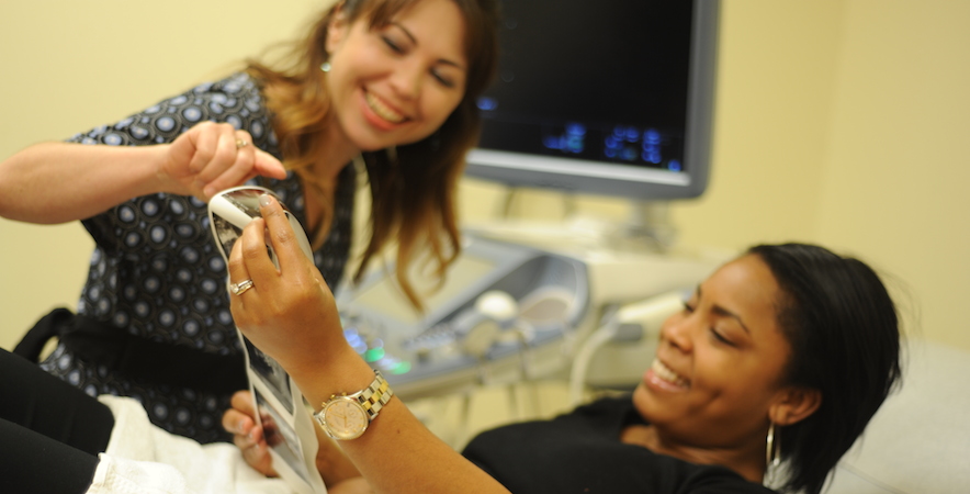
Dominion Women’s Health offers ultrasound at three of our locations. Our ultrasounds are performed by specially trained, skilled ultrasound technicians, called sonographers. You often hear the terms “ultrasound” and “sonogram.” What’s the difference? Ultrasound is the technology and sonogram is the exam itself. Usually when you’re pregnant and talking about ultrasounds, more often than not you’re really referring to sonograms.
Ultrasound imaging is also frequently used to investigate, diagnose and monitor gynecological problems such as a suspicious mass or unusual bleeding.
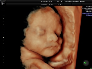
While sonographers can see the images during your exam, your physician is the one who talks about the results with you. You may be able to watch the images on a monitor during the exam however it may not be possible for you to obtain printed pictures. During pregnancy, ultrasounds will be performed periodically as determined by your doctor. Typically, you will have one performed at 20 weeks that will allow the sonographers to obtain images and measurements of your baby’s anatomy. Between 27-31 weeks, you can schedule a 3D/4D ultrasound to get a glimpse of what your baby will actually look like! Since this is not needed by your doctor, it will not be covered under your insurance. Please contact our office to learn about pricing and pre-appointment requirements. You don’t want to miss out on this moment!
How Ultrasound Works
Ultrasound works by using a “wand” or transducer to send sound waves through the body. The waves bounce back as they hit organs, tissues, and bones to create a visual image. There are two ways ultrasounds are performed:
- Transabdominal. Your abdomen is exposed, a gel is applied to the surface, and then the transducer is moved over your abdomen. For this exam, wear loose-fitting separates. You may be asked to drink and retain several glasses of water two hours before your exam. A full bladder gives us a better picture from the transabdominal exam.
- Transvaginal. A wand-shaped transducer is fitted with a condom-like latex covering, it’s lubricated, then inserted into the vagina. You change into a gown and lie in on a bed with stirrups, much like the one you have your regular exam on. Because the image is taken inside, a full bladder isn’t necessary for this ultrasound.
During Pregnancy
Ultrasound is used to see how the fetus is developing and to check for potential problems. It also gives us information about things like fetal position, the age of the fetus, and how many there are. You may have at least one general sonogram during your pregnancy.
2D, 3D, and 4D ultrasounds
 There are three types of images available through ultrasound:
- 2D is 2-dimensional meaning the images show length and width but not depth. It’s a regular black and white image. For pregnancy, it shows an outline but it may be hard for you to see the baby.
- 3D is 3-dimensional meaning the images show length, width, and depth. The technician takes several 2D images at various angles then technology translates them into a 3D image. For pregnancy, 3D shows baby’s feet, hands, and other areas the sonographer focuses on.
- 4D is 4-dimensional meaning the images show length, width, depth, and depth-over-time to show motion. This is similar to 3D but it also shows movement and can be viewed as a video. For pregnancy, 4D shows movement like thumb-sucking. 4D is rarely needed for medical reasons and is not covered by insurance.